Radiographs give clinicians a view that a visual exam alone cannot provide. For children, whose mouths are changing rapidly, small issues can evolve quickly; an X-ray often reveals hidden cavities, early infections, or unerupted teeth before symptoms appear. Identifying these concerns early makes treatment simpler, preserves healthy tooth structure, and reduces the likelihood of more invasive procedures later.
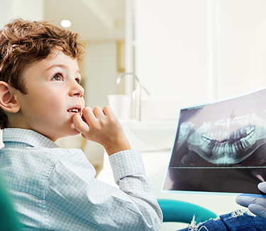
Digital systems produce high-resolution images almost instantly, which shortens appointments and helps explain findings to parents on the spot. When caregivers can see clear images and a clinician can point to a specific concern, decisions about next steps are more transparent and less stressful. That immediacy is especially valuable with young patients who benefit from efficient visits and calm, decisive care.
Beyond diagnosing current problems, radiographs are an important monitoring tool. They document eruption patterns, root development, and jaw relationships so clinicians can anticipate growth-related issues. In this way, X-rays support both immediate treatment and long-term planning—helping clinicians time interventions like space maintenance or orthodontic referrals for the best outcome.
Digital radiography uses electronic sensors to capture X-ray information and convert it into a digital image that can be enhanced and magnified. These images let clinicians examine tooth surfaces, root structures, and surrounding bone in ways that are impossible with a visual exam alone. Tiny areas of decay between teeth, the beginning of an abscess, or a fracture line can all be detected earlier with high-quality imaging.
One of the practical advantages of digital files is their adjustability: brightness, contrast, and zoom can be modified to highlight subtle findings without additional exposures. Clinicians can compare images side-by-side, mark areas of concern, and store them in the patient record for future comparison. That continuity makes it easier to identify small changes over months or years.
Digital images are also simple to share securely with other members of a child’s care team when collaborative input is needed. Whether consulting with an orthodontist about developing bite relationships or an oral surgeon about an unusual finding, high-quality images support clearer communication and more coordinated care.
Understandably, parents often worry about radiation. Modern dental radiography uses much less radiation than older film methods, and pediatric offices follow rigorous protocols to keep exposure as low as possible. Protective measures such as lead aprons and thyroid collars remain standard, and clinicians adhere to the ALARA principle—keeping doses “as low as reasonably achievable” while obtaining diagnostic-quality images.
Decisions to take X-rays are individualized. Dentists weigh the diagnostic benefit against the small exposure, taking images only when the information gained will affect diagnosis or treatment. For routine checkups, radiograph frequency is based on a child’s age, cavity risk, and dental history—not on a set schedule for every patient.
Equipment and technique also matter. Newer digital sensors require lower doses than traditional film, and trained pediatric teams use positioning aids and focused exposures to minimize repeat images. If you have questions about safety, your child’s dental team can explain why a particular view is recommended and how they limit radiation while still protecting oral health.
Bitewing radiographs are the most common images used for routine cavity detection; they show the upper and lower back teeth in one view and are particularly good at revealing decay between teeth. These quick, targeted images are frequently used for school-age children and older primary teeth where interproximal decay is harder to spot visually.
Periapical radiographs capture the entire tooth from crown to root and are used when a problem beneath the gum line is suspected—such as an infected root, a fracture, or a developmental anomaly. Panoramic images provide a single wide view of the upper and lower jaws and are helpful for evaluating overall growth, tooth eruption, and the position of permanent teeth that are still developing.
Advanced three-dimensional imaging (cone beam) is used selectively when complex anatomical detail is required for treatment planning. Because it delivers a larger dose than standard views, clinicians reserve it for situations where the added information will significantly affect care decisions—such as evaluating certain surgical or orthodontic cases.
Radiographs refine diagnosis in ways that change how clinicians treat problems. Detecting decay early often allows for more conservative management—removing less tooth structure and using preventive treatments that arrest progression. When development is closely monitored, timely interventions like space maintainers or early orthodontic guidance can prevent more complicated issues in the future.
Imaging also helps assess supporting structures such as bone height and root development, which is important for evaluating tooth stability and planning restorative work. Knowing the exact extent of decay or the relationship of a tooth to surrounding anatomy enables the team to choose the least invasive, most durable solution tailored to each child.
Finally, radiographs enhance caregiver education. Visual evidence makes preventive recommendations clearer—whether that means reinforcing brushing and flossing techniques, discussing fluoride or sealant options, or setting a schedule for monitoring. When families understand what the images show and why a particular plan is advised, they can participate confidently in decisions that support long-term oral health.
At Tiny Tots Dental Care, we use digital radiography thoughtfully—only when the information will improve diagnosis or treatment—and we prioritize safety, comfort, and clear communication. If you’d like to learn more about how radiographs are used in pediatric dentistry or whether imaging is appropriate for your child, please contact us for more information.


Email: